Cavernous Hemangioma
Orbital cavernous hemangioma is the among the most common benign neoplasm found within the adult orbit. It is a slow-growing, benign tumour involving vascular structures within the muscle cone of the orbit, which pushes the eyeball forward as it grows, resulting in proptosis. Bilateral cases are rare. It is most commonly reported in middle-age adults (20-40 years), with women more affected than men.
Symptoms of Cavernous Hemangioma
Cavernous hemangioma of the orbit presents with mass effect due to an increase in volume of the orbital contents. It causes the following symptoms and signs:
- Painless, slowly progressive bulging of the globe
- Decrease in visual acuity and visual field defects due to mass effect or involvement of the optic nerve, extraocular muscles or surrounding vasculature.
- Double vision or diplopia due to extraocular muscle dysfunction or orbital axis mismatch between the two eyes.
- Lagophthalmos due to extraocular muscle dysfunction or nerve involvement results in exposure keratopathy, keratitis, and corneal perforation.
- Pupillary dysfunction due to involvement of neural structures within the orbit.
Diagnosis of Cavernous Hemangioma
A thorough ophthalmologic examination is key to formulating an exhaustive list of differential diagnoses.
- A detailed history and review of symptoms is of paramount importance.
- Examination should be thorough and should include observation and palpation of affected eye. Hertel exophthalmometry is done to document axial proptosis.
- Assess near vision, distant vision, colour vision and visual fields followed by testing of pupillary and extraocular muscle function. Defect in any of these parameters signal compression of the optic nerve and imaging studies should be done. Extraocular muscle dysfunction is measured using prismatic evaluation.
- Slit lamp or penlight evaluation might or might not detect any abnormalities.
- Dilated fundoscopy might show choroidal folds due to mass effect while optic nerve compression shows up as visible edema, elevation, pallor, or even atrophic changes in the fundus.
Cavernous hemangioma is suspected clinically and usually confirmed with orbital imaging studies. Following investigations help to evaluate the presence and extent of the disease:
- CT scan detects an oval or round, homogenous mass with sharp margins, but falls short of a definitive diagnosis.
- A-Scan Ultrasonography shows a uniform high-echogenicity while Doppler flow study reveals decreased blood flow within the lesion.
- MRI of the cavernous hemangioma exhibits a homogenous signal. Gadolinium shows an initial central patch enhancement followed by total homogenous enhancement.
Imaging of Cavernous Hemangioma
- CT: smooth discrete lesion, fills with dye after 20 min; coronal cuts important to know tumor position relative to optic nerve. for sugical plan
- MRI: hypointense to fat on T1, hyperintense to fat on T2
- U/S: high reflectivity (A-scan high amplitude internal echoes)
Treatment of Cavernous Hemangioma
- Most cavernous hemangiomas require no treatment. The surgical approach, when indicated, depends on location and size of the tumour.
- Cavernous hemangioma involving the anterior two-thirds of the orbit is resected via an anterior eyelid, transconjunctival or transcaruncular approach. A lateral orbitotomy or its variant is more appropriate for tumours located more posteriorly. Lesions involving the orbital apex warrants a transcranial approach.
- A cryoprobe aids removal of well-circumscribed tumours with minimal risk of capsular rupture or blood loss. Carbon dioxide laser, Nd:YAG laser and Gamma knife surgery are newer modalities of treatment that can be considered.
- The visual prognosis with complete excision is excellent but incompletely excised lesions are notorious for recurrences. Occasionally, visual loss can occur as a complication of surgery.
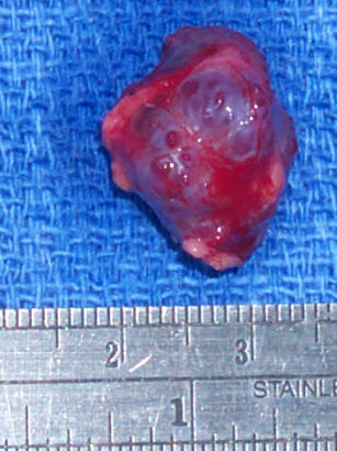
Pathology of Cavernous Hemangioma
- well encapsulated and tolerated
- shows large cavernous spaces with red blood cells